三维标本与切缘标注3D specimen and margin labels
扫描切除标本,在病理流程中记录需要再次定位的切缘位置。Scan the resected specimen and annotate margin locations identified through the pathology workflow.
针对头颈肿瘤再切除中的定位难题,将三维标本上的切缘标注经形变配准映射回创面,再通过增强现实显示目标位置。For head and neck tumor re-resection, deformable registration maps margin annotations from a scanned 3D specimen back to the wound bed, where AR displays the target location.
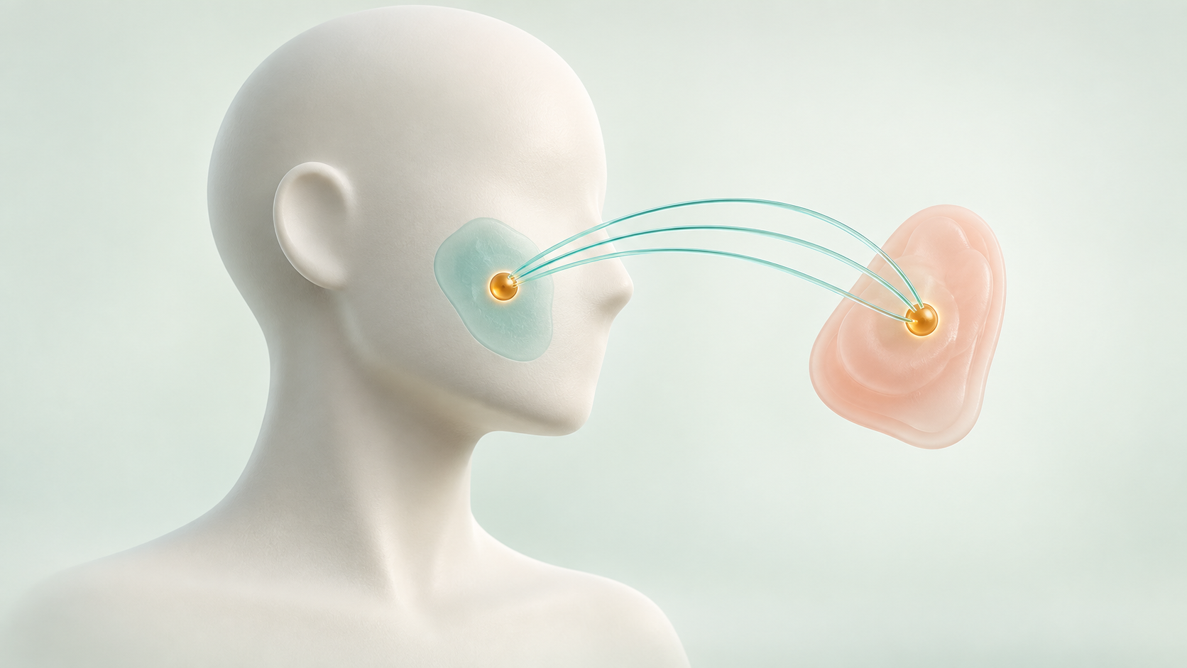
头颈肿瘤切除后,标本进入病理流程,患者的创面则留在手术台上。如果病理发现某处切缘阳性,医生需要把标本上的这个位置准确找回到创面,决定从哪里追加切除。问题在于,标本离体后会旋转、收缩和变形,它已无法像一块拼图一样直接放回原位。After a head and neck tumor is removed, the specimen enters the pathology workflow while the wound remains on the operating table. If a margin is positive, the surgeon must find its counterpart on the wound and decide where to re-resect. Rotation, shrinkage, and deformation mean the specimen no longer fits back like an unchanged puzzle piece.
“靠近上缘”或“某个方向的一角”这样的口头描述,仍需要医生结合记忆、解剖特征和标本方向进行判断。头颈区域的神经、血管和重要外观结构相邻,追加切除既要对准可疑位置,也要谨慎保留周围组织。A verbal description such as “near the upper edge” still depends on the surgeon’s memory of anatomy and specimen orientation. In the head and neck, nerves, vessels, and structures important to appearance sit close together, so additional resection must target the suspected site while carefully preserving surrounding tissue.
系统保留三维标本及其切缘标注,通过轮廓约束形变和创面配准,将目标映射回患者一侧,并锚定在 AR 视野中。病理负责判断切缘,导航负责把这一判断所对应的空间位置传递回医生眼前。The system preserves a 3D specimen with margin annotations, then uses contour-constrained deformation and wound-bed registration to map targets back to the patient and anchor them in AR. Pathology determines margin status; navigation communicates the corresponding spatial location back to the surgeon.
病理提供位置标注Pathology provides the annotation
旋转、收缩改变对应关系Rotation and shrinkage change the mapping
形变映射后原位标示Deform and map the annotation in situ
扫描切除标本,在病理流程中记录需要再次定位的切缘位置。Scan the resected specimen and annotate margin locations identified through the pathology workflow.
结合珠状缝线对应点与创面轮廓,使用 Kelvinlet 模型补偿离体形变。Bead-suture correspondences and wound contours constrain a Kelvinlet model of specimen deformation.
将形变标本配准到创面点云,并在铺巾条件下完成创面与头显坐标对齐。Register the deformed specimen to the bed point cloud, then align the bed with the headset while accounting for drapes.
将标注锚定在创面对应位置,帮助医生定位拟再次切除的区域。Anchor margin labels to the corresponding wound-bed locations to guide target relocation.
“无标记”指患者与头显之间的 AR 融合无需外部标志物;标本形变建模仍使用珠状缝线对应点,病理判断仍由病理流程提供。Marker-free refers to patient-to-headset AR fusion. Specimen deformation still uses bead-suture correspondences, and margin assessment remains part of the pathology workflow.
AR 引导端到端定位误差End-to-end localization error with AR
口头引导对照误差Error with verbal guidance
在线融合时间Online fusion time
研究在面颊与头皮两个尸体模型部位开展,包含 5 位外科住院医师及专科培训医师。口头引导加标本查看组的定位误差为 16.09 ± 4.30 mm。结果支持可行性,尚不代表临床疗效;在线融合时间不包含标本扫描等准备步骤,原型的附加手动步骤约需 10 分钟。The study included cheek and scalp cadaveric sites and five surgical residents and fellows. Verbal guidance with specimen examination yielded 16.09 ± 4.30 mm localization error. Results support feasibility, not clinical efficacy. Online fusion time excludes specimen scanning and other preparation; added manual steps took approximately 10 minutes in the prototype.
Yue Yang, Annie Benson, Matthieu Chabanas, Jason Slagle, Thomas Myles, Matthew Weinger, Jon Heiselman, Michael Miga, Michael Topf, Jie Ying Wu
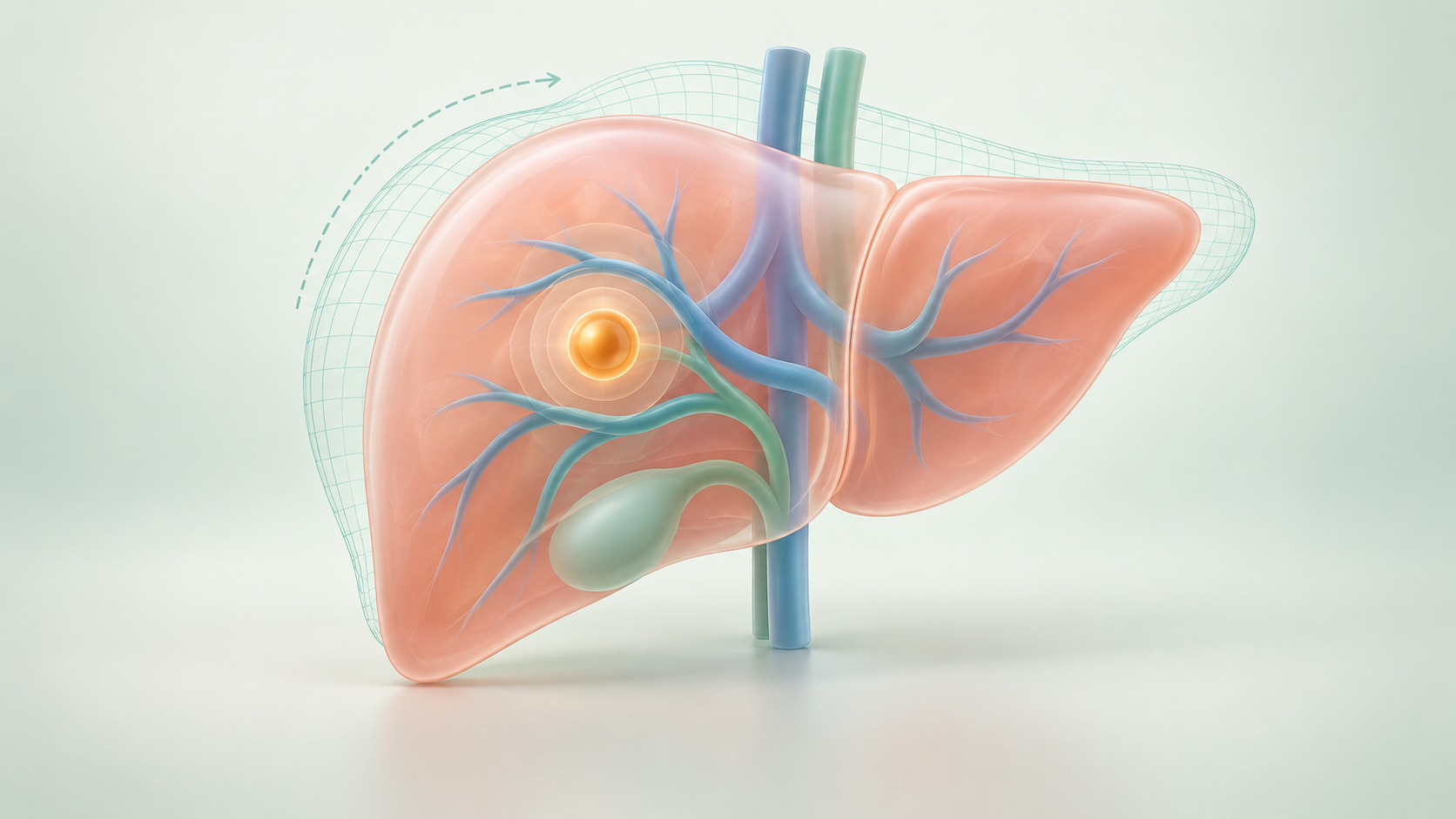
面向开腹肝脏手术,利用头显自身的双目相机重建暴露表面,自动识别解剖特征,通过生物力学形变矫正将肿瘤与血管模型叠加到术野。For open hepatic surgery, headset stereo cameras reconstruct the exposed surface. Learned anatomical features guide biomechanical correction to overlay tumor and vessel models in the operative field.
了解导航系统 →Explore the system →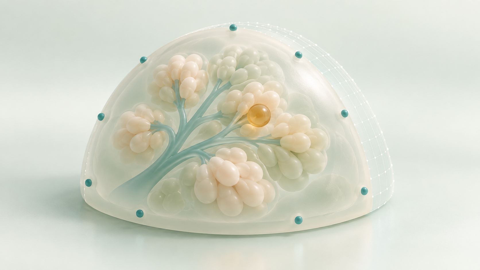
面向保乳手术,将皮肤标志点识别、乳房表面采集与 MR 显示集中在同一头显,利用非刚性配准建立术前模型与术中乳房之间的空间对应。For breast-conserving surgery, a single headset combines skin-fiducial sensing, surface acquisition, and MR display. Nonrigid registration relates the preoperative model to the intraoperative breast.
了解导航系统 →Explore the system →针对头颈肿瘤再切除中的定位难题,将三维标本上的切缘标注经形变配准映射回创面,再通过增强现实显示目标位置。For head and neck tumor re-resection, deformable registration maps margin annotations from a scanned 3D specimen back to the wound bed, where AR displays the target location.
了解导航系统 →Explore the system →